


An innovative approach to achieving full wound closure using negative pressure wound therapy and allogeneic keratinocytes
Rosemary Rose, RGN, Tissue Viability Clinical Nurse Specialist, Walsall Healthcare NHS Trust
For the purposes of this article, prior knowledge of the stages of wound healing, the benefits and application of NPWT is assumed and will not be discussed in any depth.
A pseudonym has been used, but the patient gave full consent for this case study and sharing of photographs.
Background
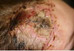
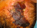
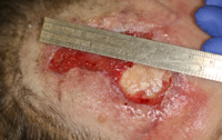
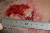
Frances, a 39 year old lady was referred to the tissue viability nurse specialist (TVNS) team to review a wound to her forehead and scalp sustained during a road traffic accident. The forehead skin flap, torn during the accident, had been reattached to the wound bed in theatre. The tissue subsequently became macerated then necrotic (Fig1, Fig 2)and required debridement in theatre under general anaesthetic (Fig 3). The wound had been debrided and dressed with both Mepitel, a non-adherent wound dressing with Safetac technology [1] to prevent adhesion or potential damage to the newly exposed tissue, and Aquacel Ag to manage exudate and as a prophylactic against infection [2]. This had been covered with a standard absorbent pad and crepe bandage to secure.
The consultant planned to apply the shark-skin graft once granulation tissue and vascular supply had been restored over the exposed bone.
Presentation
She was referred to the tissue viability (TV) service to confirm whether or not negative pressure wound therapy (NPWT) would be appropriate to manage and prepare the wound for a shark-skin graft.
Tissue Viability considerations for treatment were:
- Exudate and pain management
- Aesthetically acceptable end result as the wound was in a prominent position
- Convenience of dressing management - the patient had a very young child who still required carrying and who might dislodge the chosen dressing and cause further injury to the wound
- Number of visits
- Time to healing
Wound assessment
The wound measured approximately 12cm long x 4.5cm wide. At the base of the wound there was an area of exposed bone approximately 4cm in diameter. The maximum depth was approximately 0.5cm. The granulation tissue surrounding the bone to be bright red and healthy, and no peri-wound maceration was noted
No pain was noted, although the patient had some discomfort during dressing changes.
Following assessment, the TVNS suggested that the skin graft may be avoided as full wound closure could be achieved with an aesthetically pleasing result using NPWT until 100% full thickness granulation was achieved over the exposed bone. After discussion with the patient, and her consultant, we agreed to use Avance Solo NPWT (Mölnlycke), a single patient use delivery system, suitable for multi week treatments (lasting up to 60 days) weighing only 400 grams.
The consultant agreed to a ‘wait and see’ approach, but planned to apply a skin graft once enough granulation was achieved over the exposed bone to provide adequate vascular supply. It would be fair to say that the consultant had reservations about how successful the NPWT would be and was cautiously reluctant to accept that a skin graft would not be required.
Treatment
The consultant was concerned that it would not be possible to obtain the essential seal required for successful NPWT beneath the dressing drape due to hair growth to the peri-wound tissue. After discussion with the Mölnylcke clinical support specialist, we used Mepiseal self-curing wound sealant beneath the drape to achieve the seal. The exposed bone area was lined with a double layer of Mepitel; while this would reduce the pressure by -10mmHg per layer, it was deemed necessary by the consultant to prevent damage to the bone. All components of this dressing regimen contained Safetac technology which reduces pain and tissue damage on removal [3].
First application
The consultant set the pressure to -60mmHg in order to provide the least suction possible until assessment at the next dressing change. The pump operation was explained to the patient and she was given telephone numbers to contact the team before her next appointment if required.
The NPWT was changed every 72 hours, or as near as possible for the duration of the treatment.
Obtaining a seal was a challenge at times, possibly due to hair re-growth, so it was necessary to shave the periwound tissue each time. The patient reported that she found the NPWT delivery system easy to manage, although initially at night she found the hum a little disturbing.
At the third dressing change, it was agreed to increase the NPWT to -100mmHg; the lining and foam were cut slightly smaller than the wound bed to promote epithelialisation.
Within three weeks the overall size of the wound was reduced to 5.5cm long x 3cm wide, with the exposed bone area reduced to 2 x 2.3cm.
Week Four
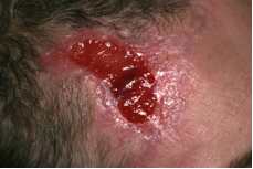
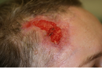
Vascularisation was observed over the bone (Fig 4, Fig 5)and it was possible to remove the double layer of mepitel with no bleeding from the granulation tissue. We therefore decided to apply it in a single layer to encourage further granulation. Foam was applied directly to the granulation tissue and the NPWT was increased to -125mmHg.
The NPWT remained at -125mmHg for three weeks. The patient had the option to reduce the pressure if she felt discomfort but this wasn’t required.
Week Seven
The overall size of the wound had reduced to approximately 5cm x 2.5cm and the exposed bone had reduced to approximately 0.3mm x 0.5mm. The NPWT was increased to -150mmHg.
Week Eight
The granulation tissue now covered 100% of the wound bed and was almost at surface skin level. NPWT was reduced to -100mmHg to prevent over granulation. The wound edges showed further epithelialisation.
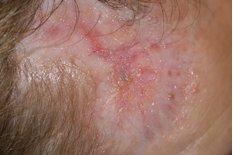
At the next dressing change the NPWT was reduced to -60mmHg as granulation was observed to be protruding through the Mepitel lining; upon removal of the Mepitel, small pale pink islands of epithelialisation were noted (Fig 6, Fig 7)
During this week over-granulation was noted (Fig 8). The MDT and the patient decided to discontinue NPWT at the next dressing change and to apply allogeneic kerarinocytes [4] in the form of CryoSkin Spray -a suspension of proliferative screened neonatal allogeneic human keratinocytes derived from a single donor [5]. As yet, it is an unlicensed medicinal product that needs to be prescribed. It is used to accelerate healing in chronic wounds and burns and different formulations have been used routinely as cultured allografts [4].
Week Nine
The CryoSkin was applied; 2ml of keratinocyte suspension was sprayed onto the wound and left for several minutes to migrate over the wound bed. It was then covered with a non-impregnated adhesive foam dressing, and a bandage to apply pressure to reduce the slight over-granulation [6].
Week Ten
An increase in exudate was noted; it was postulated that this was the suspension fluid, no as no wound deterioration nor periwound maceration was noted. CryoSkin Spray was administered.
An appointment was made to review the wound four days later as due to the previous increase in exudate, the MDT were keen to prevent maceration.
At this review, 4 x 1.5cm of exposed granulation tissue was noted at the base of the wound and a new area of exposed granulation measuring 2 x 1cm noted on the top half of the wound bed. No exudate was observed on the removed dressing nor was there any maceration to the periwound tissue. The wound was redressed with a non-impregnated adhesive foam dressing.
Week Eleven
CryoSkin was applied to both areas of granulation and redressed as before. No exudate or maceration was observed. A thin layer of epithelial tissue had migrated across 90% of the top half of the wound; however, the bottom half of the wound was overgranulating.
The TVN decided that a silver hydrofibre would be beneficial to reduce the overgranulation [7]. Mepitel was thus used on the new epithelial tissue for protection and Aquacel Ag on the overgranulation to absorb exudate and reduce potential bacterial load. Cavilon film barrier was applied to the periwound tissue and covered with an adhesive foam dressing.
The wound was reviewed three days later. The overgranulation had reduced to surface skin level, no inflammation, heat, exudate or maceration was noted. The top half of the wound was once again covered in 100% epithelial tissue. The wound was redressed with an adhesive foam dressing. For the next two weeks the dressing were changed twice per week. Epithelial migration continued across the wound bed.
Week 13
The exposed granulation area had reduced to 1.2cm x 1cm no further signs of overgranulation were noted. Ccryoskin was applied and the wound covered with an adhesive non impregnated foam dressing.
Four days later 100% epithelialisation was observed. Mepitel was applied to the new tissue with an adhesive foam for protection. The patient redressed the wound for the next three weeks.
Weeks 16
Mepiform Self-adherent soft silicone dressing was applied to reduce the potential scarring to the new tissue. This proved to be more acceptable and no further tissue was removed.
Following this, the wound was reviewed periodically by the consultant, no further problems ensued. The patient kindly sent me a photograph of the wound taken seven months later (Fig 9)
Discussion
This wound was effectively closed in full by secondary intention with the application of NPWT and allogeneic keratinocytes within five months. The need for an allograft and surgery was eliminated by implementing this cost effective alternative.
The aesthetic appearance of the wound was acceptable to the patient, with minimal scarring and colour very close to that of the original forehead tissue.
The use of the Avance Solo NPWT system provides all the advantages of other NPWT systems with the added benefits of allowing mobility and flexibility. Patient’s using the Avance Solo are able to easily maintain their normal activities of daily living with minimal impact due to the compact size of the pump casing and the ease with which it can be carried and hidden. Considering these patient needs meets the demands of the NHS White Paper [8] and Augustin et al [9]. Thus, this treatment should be explored and utilised where healing requiring minimal scarring is key and where surgery can be avoided with minimal discomfort to the patient.
Conclusion
Compared to the costs that surgery and the subsequent follow up that would have been required, this treatment plan was cost efficient. It is vital that TVN’s research and implement cost effective treatments that both improve patient care and raise the profile of their speciality [10]. Nursing metrics provide useful and meaningful information to measure successes and changes in practice which will improve patient outcomes . In this case the patient outcomes were considered highly successful.